Create articles from any YouTube video or use our API to get YouTube transcriptions
Start for freeEngaging with Eye Anatomy Through Questions and Answers
Learning about the anatomy of the eye can be a fascinating journey, especially when approached through an interactive, question-based format. This method not only makes the learning process engaging but also helps in retaining information effectively.
Understanding the Basic Structures of the Eye
The human eye is composed of various structures, each playing a crucial role in how we perceive the world. The cornea and sclera are parts of what is known as the fibrous tunic. The cornea is clear and allows light to enter the eye, while the sclera, being white, provides protection and form.
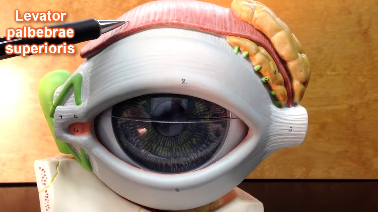
Muscles and Nerves Controlling Eye Movements
Eye movements are controlled by several muscles which are, in turn, regulated by nerves. The superior rectus and medial rectus are two such muscles. It's interesting to note that while most of these muscles are controlled by the oculomotor nerve, exceptions like the superior oblique and lateral rectus are controlled by trochlear and abducens nerves respectively.
Layers Beyond What Meets the Eye
Deeper within these external structures lie more intricate layers like the vascular tunic, which includes both the ciliary body and choroid. These play roles in maintaining proper vision function by regulating focus and nourishing other parts of the eye.
The Role of Iris, Pupil, and Lens in Vision
The colored part of your eye is known as the iris, with a central opening called the pupil—essentially a hole through which light enters. Behind this lies the lens, crucial for focusing light onto your retina.
Retina's Composition for Optimal Vision
The retina itself consists of two layers - an outer pigmented layer that absorbs excess light to prevent confusion in vision, and an inner neural layer that processes visual information. Notably, specific areas like the macula lutea concentrate on high amounts of cones for sharp central vision while areas like optic disc act as blind spots where no image detection occurs.
Additional Eye Components Supporting Vision
Other components include lacrimal glands responsible for tear production; lacrimal puncta involved in tear drainage; conjunctiva that can become inflamed during conjunctivitis (pink eye); along with nerves like trochlear controlling specific muscles such as Superior oblique.
Crossroads at Optic Chiasma
A critical point within our visual pathway is where optic nerves cross at what's known as optic chiasma. Here signals from both eyes integrate before transmitting to brain regions responsible for processing visual cues. This crossing ensures that visual information from both eyes is combined effectively for a coherent field of view.
Engage Actively with Your Learning Process
This interactive approach not only clarifies complex concepts but also encourages you to actively engage with your learning process. By asking questions first then revealing answers later encourages critical thinking skills necessary not just for academic success but lifelong learning too.
Article created from: https://youtu.be/XGnAZA-Uc9I