Create articles from any YouTube video or use our API to get YouTube transcriptions
Start for freeThe Complex World Inside Our Eyes
The human eye is a marvel of biological engineering. Each component plays a critical role in how we perceive the world. This detailed overview will guide you through various structures of the eye and their functions, enhancing your understanding of this vital organ.
The Journey of a Tear
Tears are essential for maintaining eye health, providing moisture and protection from irritants. The lacrimal gland, located at the upper outer region of each eye, is responsible for tear production. Tears flow from here through tiny ducts across the eye towards the medial canthus, eventually draining down through the nasolacrimal duct to the nasal cavity. This connection explains why our nose runs when we cry.
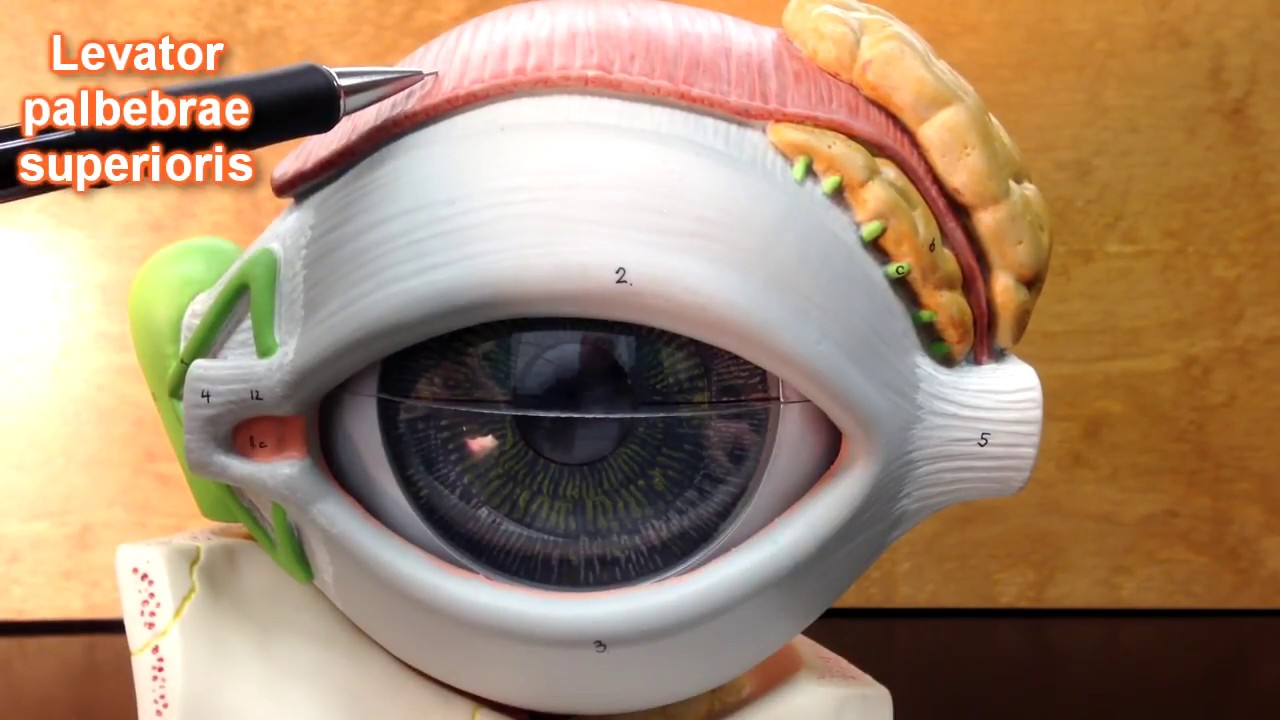
Muscles that Move and Protect
Several muscles around the eye play roles in movement and protection. The levator palpebrae superioris muscle is particularly important as it controls the lifting of the upper eyelid, enabling us to open our eyes. Surrounding muscles like superior rectus, inferior rectus, lateral rectus, and medial rectus assist in moving the eyeball in different directions, providing us with a broad field of view.
Focusing on Vision - Cornea and Lens
The front part of our eyes features two critical components for focusing light — the cornea and the lens. The cornea acts as a powerful light bender; when light passes through it gets directed onto our retina at the back of our eyes. For those who wear glasses or contact lenses, these adjustments help compensate for any focusing issues originally present in how their cornea bends light.
The lens sits just behind; it fine-tunes focus allowing us to see details clearly both up close and far away. Supported by suspensory ligaments connected to ciliary muscles, its shape can change—thicker for closer objects and thinner for distant ones—aiding in dynamic focusing.
Internal Eye Dynamics - Iris and Pupil Regulation
Deep within our eyes lies an adjustable aperture known as the pupil, controlled by muscles within colored part known as the iris which gives us our unique eye colors. Depending on lighting conditions or emotional states, this pupil can widen or constrict to regulate how much light enters further inside.
Supporting Structures - Choroid Layer & Vitreous Humor
Beneath these lie supportive layers that play crucial roles too; one such layer is the choroid layer filled with blood vessels supplying nutrients and oxygen vital for maintaining retinal health. Behind all this lies a gel-like substance called vitreous humor filling most volume inside eyeball helping maintain its shape while also protecting retina by absorbing excess heat generated by incoming light rays.
Retina - The Ultimate Image Processor
At back surface inside lies retina; an intricate layer packed with millions photoreceptors called rods cones detecting colors low-light conditions respectively center part called macula has highest concentration cones especially fovea centralis where sharpest images formed due direct focus incoming rays here making it crucial reading recognizing faces other detailed activities.
In conclusion understanding anatomy functionality each part helps appreciate complexity behind simple act seeing encourages care protect precious sense sight.
Article created from: https://youtu.be/JuHoqmDBGoM


