Create articles from any YouTube video or use our API to get YouTube transcriptions
Start for freeWelcome to an educational journey through the human skull, an essential segment of anatomy that houses the brain and forms the structure of the face. This guide will navigate through the various bones, foramina, and other critical structures that make up the skull, elucidating their functions and significance in human anatomy. For more enlightening resources on anatomy and physiology, be sure to explore humanbodyhelp.com.
The Skull Without the Mandible
By removing the mandible, we gain a clearer view of the interior structures of the skull. This perspective reveals:
- Maxillary Bone: Notably, the palatine process of the maxillary bone.
- Palatine Bones: The horizontal plates forming the hard palate.
- Pterygoid Processes: Including both the lateral and medial pterygoid plates.
- Vomer Bone: Situated centrally within the skull.
One intriguing feature is a hole known as the greater palatine foramen, which facilitates the passage of the greater palatine nerve.
Ethmoid Bone and Nasal Conchae
Tilting the skull reveals the ethmoid bone and the inferior nasal conchae—critical components of the nasal structure. Above these, although seemingly below due to the skull's inversion in this view, are the middle and superior nasal conchae.
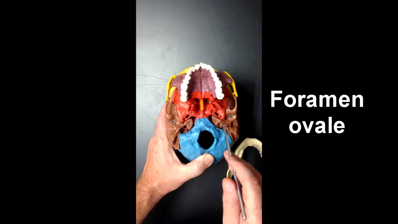
The Foramina of the Skull Base
The skull's base is punctuated by several foramina (holes), each serving as a passage for nerves or vessels:
- Foramen Ovale: A bilateral oval opening for the mandibular branch of the trigeminal nerve.
- Foramen Spinosum: Allows passage for the internal meningeal artery.
- Foramen Lacerum: Positioned between the sphenoid, temporal, and occipital bones, it permits the transit of various structures, including the artery and nerve of the pterygoid canal, and the ascending pharyngeal artery.
- Carotid Canal: Through which the internal carotid artery enters the skull to supply blood to the brain.
- Jugular Foramen: For the exit of the internal jugular vein.
- Stylomastoid Foramen: Enables the facial nerve to exit the skull.
Additionally, the hypoglossal canal allows for the passage of the hypoglossal nerve (cranial nerve XII), and the condyloid foramen provides a route for occipital emissary veins.
Foramen Magnum and Mastoid Notch
The foramen magnum, a large opening at the skull's base, is crucial for the passage of the spinal cord. Adjacent to this, the mastoid notch, which is not a foramen but a notable structure, serves as an anchor for one of the suprahyoid muscles.
Conclusion
The skull's anatomy is a complex and fascinating subject, integral to understanding how the human body protects its most vital organ—the brain—and facilitates various functions through the passages provided by its foramina. Whether you're a student, educator, or simply an anatomy enthusiast, diving into the specifics of skull anatomy reveals the incredible precision and functionality designed into the human body.
If you've found this exploration insightful, consider subscribing for more educational content and don't hesitate to visit humanbodyhelp.com for additional resources.


